Management
Types of Intervention
Treatment of ankle syndesmosis injuries is broadly divided into surgical and non-surgical/conservative (20). Surgery is generally indicated when there are large amounts of instability in the ankle, or a large diastasis between tibia and fibula (21).
Surgery involves one or two screws through both the tibia and fibula, 2-3cm above the talocrural joint, crossing the syndesmosis (22). Screw type and size appears to vary based on the preferences of the orthopaedic surgeon (20). Screws do not remain long term, and are removed; however there are variations in timeframe for screw removal which will be discussed further.
The process of rehabilitation is generally the similar if not the same in both surgical and non-surgical treatments. It is, however, important to note that non-surgical interventions generally result in a longer duration of the rehabilitation process (23).
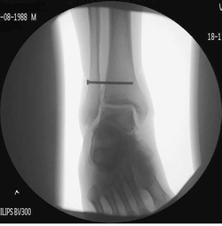
Figure 11: An example of fixation using a 3.5mm screw (20).
Rehabilitation
Although current literature does not outline a specific rehabilitation protocol for ankle syndesmosis injuries, most research indicates that a three staged approach, with specific goals per stage, is the preferred strategy for most practitioners. The stages typically consist of acute management, sub-acute management, and advanced training (the latter is primarily for athletes). It is suggested that the progression through stages should be dependent on the individual patient (4), and movement between stages may be indicated by the achievement of functional goals rather than time-specific parameters (22).
Stage 1: Acute
Goal: Protect the injury and minimise inflammation (23).
Weight bearing status: Weight bearing immediately post injury, and up 2 weeks post injury should be limited to protect the healing structures, however will vary between individuals. Patients with poor muscle control, or a severe injury with associated fractures may be completely immobilised in a cast (21). Patients requiring internal fixation surgery may also have restricted weight bearing for up to two weeks (23) however there is controversy in this area. It has been suggested that weight bearing should not begin until fixation screws have been removed to minimise long term effects on bone and the interosseous membrane (20), however recent research using college athletes has suggested that early weight bearing, well within the two week period post-surgery, has produced comparable long term outcomes to typical interventions, with the benefit of early return to participation in sport and no interruption in training, with screw removal occurring up to 8-12 weeks post-surgery (9).
Immobilisation: Level of immobilisation is dependent on the severity of the injury, and it is the role of the physiotherapist to ascertain the history of the injury based on medical imaging and observations. Unstable syndesmosis injuries, particularly with a large tibia-fibula diastasis, should be completely immobilised (21), however immobilisation may be decreased in less severe injuries, with greater stability, and support can be achieved through the use of a brace or stirrup (23), while still minimising end range dorsiflexion and external rotation (4).
Ambulation: It is unlikely that patients will be able to ambulate without the assistance of a walking aid such as crutches (23). Attempts at ambulation may begin in the acute stage, and as the therapist it is important to allow motion into dorsiflexion provided it is not end range (4). An exercise management program should be designed by the therapist to progress walking from flat surfaces to stairs and uneven surfaces, beginning with walking in a straight line, and progressing to direction changes and increased ambulation speed up to jogging (4).
Stage 2: Subacute
Goal: Increase mobility, normalise movement, improve strength.
Immobilisation/mobilisation: The ankle joint should no longer be immobilised at this stage, which may be up to 4 weeks post injury/surgical intervention, and any casts or boots should be removed provided the patient is pain-free in the resting joint (22). At this stage patient and therapist should aim to achieve close to full range of motion via long stretching of plantar-flexor, inversion and eversion muscles, combined with manual joint mobilisations, (anterior-posterior, inversion-eversion) while still avoiding end range external rotation and dorsiflexion (23).
Strength training: Strength of muscles around the ankle joint contribute to joint stability, and will naturally increase as proficiency in basic tasks, such as walking and jogging increase (4). A strength training protocol should be designed by the therapist to suit the patient, and should progress gradually. It is suggested the use of resistance bands initially is appropriate to increase strength in dorsiflexion, plantar flexion and supination/pronation, which may then progress to the use of weights, including body weight such as during calf-raises (4). It is also essential to facilitate neural mechanisms, and return reflexes and balance in the affected joint. Training aids such as wobble boards may be used to promote improved proprioception and reflexes, progressing from double leg to single leg standing and additional functional tasks such as squatting. The use of manual muscle testing may be used to establish strength goals, using the unaffected ankle as a model, and these strength measures can be used for functional reassessment of strength improvements as rehabilitation progresses (24).
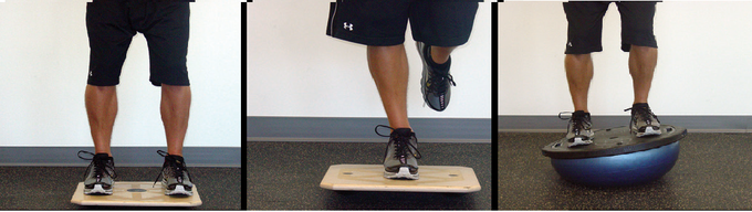
Figure 12: Progression of ankle strength exercises; double leg on balance board, single leg on balance board, double leg on wobble board (23).
Stage 3: Advanced Training
Goal:To prepare any injured athletes for return to normal activity.
Weight bearing/ambulation: Should all be normal by this stage of rehabilitation.
Training: Training should now begin in sports specific tasks. This will be different dependent on the sport, but in most cases pain free running, jumping and hopping, as well as side to side movement, should be achievable (23). Proprioceptive taping, and the use of ankle braces may still be employed for better stability outcomes (21). The main role of the therapist at this stage is to ensure the quality of the movement, noting any deficiencies, investigating any sources of pain, and observing stability (23).
