Anatomy, Signs and Symptoms
Sprains to the ligaments of the ankle joint are an extremely common injury sustained by both the sedentary and athletic population, with lateral ligament sprains actually rated as the most frequently injured site in the human body (1). (2) found the lateral ligament complex to constitute 77%, the medial complex 14% and isolated syndesmosis injuries accounting for five percent of all ankle sprains. However, a more recent study (3) has demonstrated that syndesmotic injuries are actually more common than other types of sprain in certain circumstances, that is; during collision sports and those like skiing and hockey in which the foot is immobilised in a boot (4).
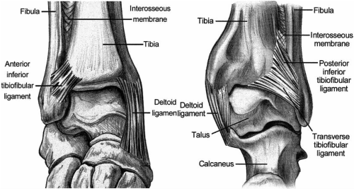
Figure 1: The anterior and posterior views of the syndesmosis of the ankle (12).
Anatomy
The tibiofibular syndesmosis, located at the distal articulation of the tibia and the fibula, comprises four fundamental structures to maintain stability and integrity at the ankle joint. These include the interosseous membrane, transverse ligament and the anterior and posterior inferior tibiofibular ligaments (AITFL and PITFL respectively) (5). A syndesmotic ankle sprain, also referred to as a high ankle sprain (6), involves damage to one or more of these sites and is most commonly sustained in conjunction with a rupture of the deltoid ligament (3) or a bony avulsion of the tibia, but can also involve fracture of the fibula (termed Maissoneuve fractures) (7).
The tibiofibular syndesmosis, located at the distal articulation of the tibia and the fibula, comprises four fundamental structures to maintain stability and integrity at the ankle joint. These include the interosseous membrane, transverse ligament and the anterior and posterior inferior tibiofibular ligaments (AITFL and PITFL respectively) (5). A syndesmotic ankle sprain, also referred to as a high ankle sprain (6), involves damage to one or more of these sites and is most commonly sustained in conjunction with a rupture of the deltoid ligament (3) or a bony avulsion of the tibia, but can also involve fracture of the fibula (termed Maissoneuve fractures) (7).
Symptoms
Differentiating this type of injury from other ankle sprains of the lateral or medial ligament complexes is integral to maximise the effectiveness of treatment and management. Clinical presentation of a typical syndesmotic injury involves symptoms localised above the tibiotalar joint, indicative of a high ankle sprain (8). Tenderness is notably more proximal than that seen in routine lateral ankle sprains, with pain over the AITFL (9), proximally over the interosseous membrane and/or over the PITFL (10). The patient will report symptoms of anterior pain over the distal part of the tibia and fibula and posterior medial ankle joint pain. They will also report ankle instability, and pain during weight bearing, passive dorsiflexion and external rotation and during toe off (10), with (11) finding instability to be the primary complaint on presentation of acute injuries with pain and functional deficiencies the main concern in chronic injuries.
Differentiating ankle sprains
The main differentiation between the potential types of ankle sprain on first presentation, is the anatomical location of the symptoms. Pain experienced over the lateral or medial ligaments are diagnostic of injury to these areas, while anterior lateral ankle pain in the vicinity of the syndesmosis is a strong indicator that this structure has been damaged (5).
Signs
While swelling and contusions may also be present with an acute injury, they are usually less prominent than that expected with a lateral sprain (another aspect that differentiates the two).
Used in combination with the history and reported mechanism of injury, these signs and symptoms will assist the diagnostician to appropriately evaluate the joint.
