Mechanism of Injury
Like most ligamentous sprains, a sprain to the tibiofibular syndesmosis is not insidious in nature, there is a clearly defined traumatic event masquerading as the mechanism of injury. However, to add ambiguity to the diagnostic process, there is no one particular movement that has been correlated to be the sole cause of syndesmotic injuries. There is evidence to some extent that suggests eversion, inversion, plantarflexion, pronation and internal rotation can all result in a syndesmotic injury if the force exerted on the joint is great enough (12). The two most commonly reported and widely accepted explanations are that syndesmosis injuries primarily occur following external rotation (9) or hyperdorsiflexion (12).
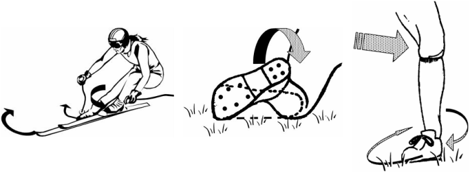
Figure 2: Potential sources of sustaining an injury to the syndesmosis (12).
External Rotation: The underlying problem with an external rotation moment around the ankle joint, is the associated widening of the mortise (the hinge that connects the distal tibia and fibula to the talus), injuring the syndesmosis in the process (12). The talus is normally positioned to allow only insignificant rotation, but it may be rotated laterally under excessive external rotation of the forefoot. This results in separation of the tibia and fibula as the fibula is pushed externally by the movement of the talus. The amount of force exerted in this direction is directly correlated with the extent of damage; ranging from tearing of the AITFL, PITFL, transverse ligament and the interosseous membrane, to full rupture of these structures, associated bony avulsions and even fibula fractures.
- This type of injury has been shown to occur in skiers (Figure 2a) and also footballers- when a blow is sustained to the lateral leg of a prone player with an externally rotated foot (Figure 2b), or when the body moves in the opposite direction to a lateral blow to the knee while the foot is fixed in external rotation (Figure 2c).
- This type of injury has been shown to occur in skiers (Figure 2a) and also footballers- when a blow is sustained to the lateral leg of a prone player with an externally rotated foot (Figure 2b), or when the body moves in the opposite direction to a lateral blow to the knee while the foot is fixed in external rotation (Figure 2c).
Hyperdorsiflexion: Forced separation of the malleoli can also occur during extreme hyperdorsiflexion by the anterior portion of the talus, which is characteristically wider than the posterior aspect. - This type of injury is usually sustained by athletes competing in running or jumping sports when momentum carries their weight forward when they come to an abrupt stop or if they fall forward on a fixed foot.
Excessive Eversion and Inversion: Rupture of the medial ligaments through excessive eversion at the subtalar joint can also result in widening of the mortise as the talus forces the fibula laterally. Extreme inversion about this joint can also separate the fibula and the tibia through lateral ligament damage (13). If the mechanism of injury originates from one of these two, the medial and lateral malleoli and head of the fibula should be imaged thoroughly for injury, as they are susceptible to fracture before syndesmotic damage is expected (14).
Internal Rotation: This injury can also be sustained when the tibia is rotated internally while a pronated and everted foot remains in a fixed position (3). This eversion component of the movement explains the medial ligament damage often associated with these injuries.
